
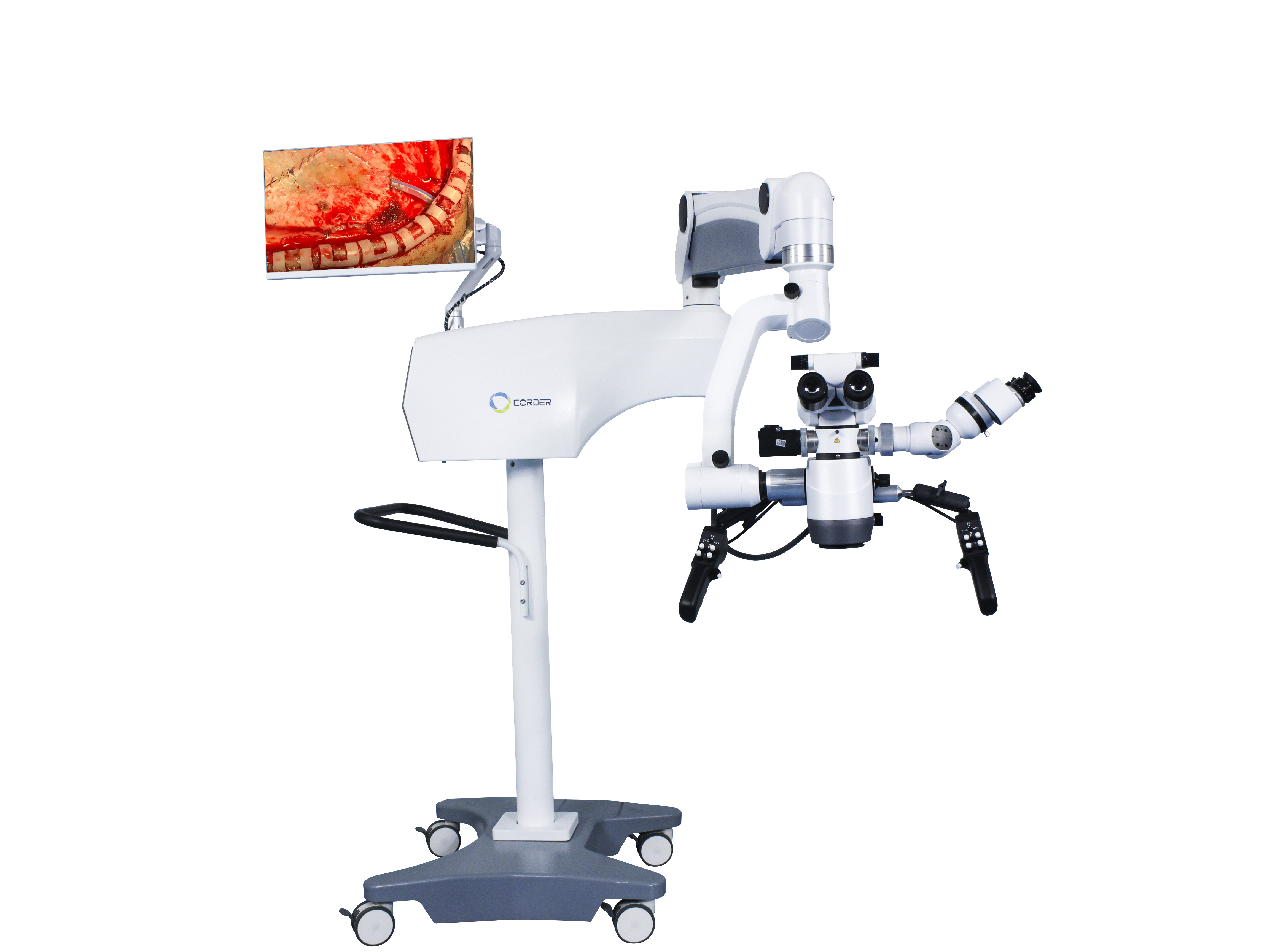
న్యూరోసర్జరీలో సర్జికల్ మైక్రోస్కోప్ల అప్లికేషన్ చరిత్ర మరియు పాత్ర
న్యూరోసర్జరీ చరిత్రలో, అనువర్తనంశస్త్రచికిత్స మైక్రోస్కోప్లుఇది ఒక అద్భుతమైన చిహ్నం, ఇది కంటితో చూస్తూ శస్త్రచికిత్స చేసే సాంప్రదాయ న్యూరోసర్జికల్ యుగం నుండి, ఒక నిర్దిష్ట పరికరం సహాయంతో శస్త్రచికిత్స చేసే ఆధునిక న్యూరోసర్జికల్ యుగానికి పురోగమిస్తోంది.సూక్ష్మదర్శినిఎవరు మరియు ఎప్పుడుఆపరేటింగ్ మైక్రోస్కోప్లున్యూరోసర్జరీలో ఉపయోగించడం ప్రారంభించారా? దీని పాత్ర ఏమిటి?శస్త్రచికిత్స మైక్రోస్కోప్న్యూరోసర్జరీ అభివృద్ధిలో పాత్ర పోషించిందా? విజ్ఞాన, సాంకేతిక రంగాల పురోగతితో,ఆపరేటింగ్ మైక్రోస్కోప్మరికొన్ని అధునాతన పరికరాలతో భర్తీ చేయాలా? ఇది ప్రతి న్యూరోసర్జన్ తెలుసుకోవలసిన ప్రశ్న, మరియు న్యూరోసర్జరీ రంగంలో సరికొత్త సాంకేతిక పరిజ్ఞానాన్ని, పరికరాలను ఉపయోగించి, న్యూరోసర్జరీ శస్త్రచికిత్సా నైపుణ్యాలను మెరుగుపరచాలి.
1. వైద్య రంగంలో మైక్రోస్కోపీ అనువర్తనాల చరిత్ర
భౌతిక శాస్త్రంలో, కళ్లద్దాల కటకాలు అనేవి ఒకే నిర్మాణంతో ఉండి, వస్తువులను పెద్దవిగా చేసే ప్రభావాన్ని కలిగి ఉండే కుంభాకార కటకాలు. వీటి ఆవర్ధనం పరిమితంగా ఉంటుంది, వీటిని భూతద్దాలు అని కూడా పిలుస్తారు. 1590లో, ఇద్దరు డచ్ ప్రజలు ఒక సన్నని స్థూపాకార గొట్టం లోపల రెండు కుంభాకార కటక ఫలకాలను అమర్చి, ప్రపంచంలోనే మొట్టమొదటి సంయుక్త నిర్మాణ భూతద్ద పరికరాన్ని ఆవిష్కరించారు: అదే...సూక్ష్మదర్శినిఆ తర్వాత, మైక్రోస్కోప్ నిర్మాణం నిరంతరం మెరుగుపరచబడింది మరియు దాని ఆవర్ధనం నిరంతరం పెరిగింది. ఆ సమయంలో, శాస్త్రవేత్తలు ప్రధానంగా దీనిని ఉపయోగించారు.సంయుక్త సూక్ష్మదర్శినిజంతువులు మరియు మొక్కల కణాల నిర్మాణం వంటి సూక్ష్మ నిర్మాణాలను పరిశీలించడానికి. 19వ శతాబ్దం మధ్య నుండి చివరి వరకు, భూతద్దాలు మరియు మైక్రోస్కోపులు క్రమంగా వైద్య రంగంలో ఉపయోగించబడ్డాయి. మొదట్లో, శస్త్రవైద్యులు శస్త్రచికిత్స కోసం ముక్కు వంతెనపై ఉంచగలిగే ఒకే కటకం నిర్మాణంతో కూడిన కళ్లద్దాల శైలి భూతద్దాలను ఉపయోగించారు. 1876లో, జర్మన్ వైద్యుడు సేమిష్ సంయుక్త కళ్లద్దాల భూతద్దాన్ని ఉపయోగించి ప్రపంచంలోనే మొట్టమొదటి "సూక్ష్మదర్శిని" శస్త్రచికిత్సను నిర్వహించారు (ఆ శస్త్రచికిత్స ఏ రకమైనదో తెలియదు). 1893లో, జర్మన్ కంపెనీ జైస్ దీనిని కనుగొంది.బైనాక్యులర్ మైక్రోస్కోప్ఇది ప్రధానంగా వైద్య ప్రయోగశాలలలో ప్రయోగాత్మక పరిశీలన కోసం, అలాగే నేత్ర వైద్య రంగంలో కార్నియల్ మరియు పూర్వ ఛాంబర్ గాయాలను పరిశీలించడానికి ఉపయోగించబడుతుంది. 1921లో, జంతువుల లోపలి చెవి శరీర నిర్మాణ శాస్త్రంపై ప్రయోగశాల పరిశోధన ఆధారంగా, స్వీడిష్ ఓటోలారింగాలజిస్ట్ నైలెన్ ఒక స్థిరమైన దానిని ఉపయోగించారు.మోనోక్యులర్ సర్జికల్ మైక్రోస్కోప్మానవులపై దీర్ఘకాలిక ఓటిటిస్ మీడియా శస్త్రచికిత్స చేయడానికి అతను స్వయంగా రూపొందించి, తయారు చేసిన ఒక నిజమైన మైక్రోసర్జరీని ప్రవేశపెట్టాడు. ఒక సంవత్సరం తరువాత, నైలెన్ యొక్క ఉన్నతాధికారి వైద్యుడు హ్లోల్మ్గ్రెన్ ఒకబైనాక్యులర్ సర్జికల్ మైక్రోస్కోప్ఆపరేటింగ్ రూమ్లో జైస్ చేత తయారు చేయబడింది.
ప్రారంభంలోఆపరేటింగ్ మైక్రోస్కోప్లుపేలవమైన యాంత్రిక స్థిరత్వం, కదలలేకపోవడం, వివిధ అక్షాలపై కాంతి ప్రసరించడం మరియు ఆబ్జెక్టివ్ లెన్స్ వేడెక్కడం, ఇరుకైన శస్త్రచికిత్స మాగ్నిఫికేషన్ క్షేత్రం మొదలైన అనేక లోపాలు ఉన్నాయి. ఇవన్నీ దీని విస్తృత అనువర్తనాన్ని పరిమితం చేసే కారణాలు.శస్త్రచికిత్స మైక్రోస్కోప్లుతరువాతి ముప్పై సంవత్సరాలలో, శస్త్రవైద్యులు మరియుమైక్రోస్కోప్ తయారీదారులు, పనితీరుశస్త్రచికిత్స మైక్రోస్కోప్లునిరంతరం మెరుగుపరచబడింది, మరియుబైనాక్యులర్ సర్జికల్ మైక్రోస్కోప్లు, పైకప్పుపై అమర్చిన మైక్రోస్కోప్లుజూమ్ లెన్సులు, కోయాక్సియల్ కాంతి మూల ప్రకాశం, ఎలక్ట్రానిక్ లేదా నీటి పీడనంతో నియంత్రించబడే కీలు చేతులు, ఫుట్ పెడల్ నియంత్రణ మొదలైనవి వరుసగా అభివృద్ధి చేయబడ్డాయి. 1953లో, జర్మన్ కంపెనీ జైస్ (Zeiss) ఒక ప్రత్యేకమైన శ్రేణిని ఉత్పత్తి చేసింది.ఓటాలజీ కోసం శస్త్రచికిత్స మైక్రోస్కోప్లుముఖ్యంగా మధ్య చెవి మరియు కణతల ఎముక వంటి లోతైన గాయాలపై చేసే శస్త్రచికిత్సలకు అనువైనది. అయితే పనితీరుశస్త్రచికిత్స మైక్రోస్కోప్లుఅభివృద్ధి కొనసాగుతున్నందున, శస్త్రవైద్యుల ఆలోచనా విధానం కూడా నిరంతరం మారుతోంది. ఉదాహరణకు, జర్మన్ వైద్యులైన జోల్నర్ మరియు వుల్స్టెయిన్ ఈ విధంగా పేర్కొన్నారుశస్త్రచికిత్స మైక్రోస్కోప్లుకర్ణభేరి ఆకృతి శస్త్రచికిత్స కోసం తప్పనిసరిగా ఉపయోగించాలి. 1950ల నుండి, నేత్ర వైద్యులు నేత్ర పరీక్షల కోసం కేవలం మైక్రోస్కోప్లను మాత్రమే ఉపయోగించే పద్ధతిని క్రమంగా మార్చి ప్రవేశపెట్టారు.ఓటోసర్జికల్ మైక్రోస్కోప్లునేత్ర శస్త్రచికిత్సలోకి. అప్పటి నుండి,ఆపరేటింగ్ మైక్రోస్కోప్ఓటాలజీ మరియు ఆప్తాల్మాలజీ రంగాలలో విస్తృతంగా ఉపయోగించబడుతున్నాయి.
2. న్యూరోసర్జరీలో సర్జికల్ మైక్రోస్కోప్ యొక్క అనువర్తనం
న్యూరోసర్జరీ యొక్క ప్రత్యేకత కారణంగా,న్యూరోసర్జరీలో శస్త్రచికిత్స మైక్రోస్కోప్లుఓటాలజీ మరియు ఆప్తాల్మాలజీ కంటే ఇది కొంచెం ఆలస్యంగా వస్తుంది, మరియు న్యూరోసర్జన్లు ఈ కొత్త సాంకేతికతను చురుకుగా నేర్చుకుంటున్నారు. ఆ సమయంలో,శస్త్రచికిత్స మైక్రోస్కోప్ల వాడకంప్రధానంగా యూరప్లో ఉండేది. అమెరికన్ నేత్ర వైద్యుడు పెరిట్ మొదట పరిచయం చేశారుశస్త్రచికిత్స మైక్రోస్కోప్లు1946లో ఐరోపా నుండి యునైటెడ్ స్టేట్స్కు, అమెరికన్ న్యూరో సర్జన్లు ఉపయోగించడానికి పునాది వేసిందిఆపరేటింగ్ మైక్రోస్కోప్లు.
మానవ జీవిత విలువను గౌరవించే దృక్కోణం నుండి, మానవ శరీరం కోసం ఉపయోగించే ఏదైనా కొత్త సాంకేతికత, పరికరాలు లేదా సాధనాలు తప్పనిసరిగా ప్రాథమిక జంతు ప్రయోగాలు మరియు ఆపరేటర్లకు సాంకేతిక శిక్షణకు లోనవ్వాలి. 1955లో, అమెరికన్ న్యూరోసర్జన్ మాలిస్ ఒక పద్ధతిని ఉపయోగించి జంతువులపై మెదడు శస్త్రచికిత్సను నిర్వహించారు.బైనాక్యులర్ సర్జికల్ మైక్రోస్కోప్అమెరికాలోని సదరన్ కాలిఫోర్నియా విశ్వవిద్యాలయంలో న్యూరో సర్జన్ అయిన కుర్జే, మైక్రోస్కోప్ కింద చెవి శస్త్రచికిత్సను గమనించిన తర్వాత, ప్రయోగశాలలో మైక్రోస్కోప్ను ఉపయోగించే శస్త్రచికిత్సా పద్ధతులను నేర్చుకోవడానికి ఒక సంవత్సరం గడిపారు. 1957 ఆగస్టులో, ఆయన ఒక 5 ఏళ్ల బాలుడికి అకౌస్టిక్ న్యూరోమా శస్త్రచికిత్సను విజయవంతంగా నిర్వహించారు.చెవి శస్త్రచికిత్స మైక్రోస్కోప్ఇది ప్రపంచంలోనే మొట్టమొదటి మైక్రోసర్జికల్ శస్త్రచికిత్స. ఆ తర్వాత కొద్దికాలానికే, కుర్జే ఒక పరికరాన్ని ఉపయోగించి ఆ చిన్నారికి ఫేషియల్ నర్వ్ సబ్లింగువల్ నర్వ్ అనస్టోమోసిస్ను విజయవంతంగా నిర్వహించారు.శస్త్రచికిత్స మైక్రోస్కోప్మరియు ఆ బిడ్డ కోలుకోవడం అద్భుతంగా జరిగింది. ప్రపంచంలో ఇది రెండవ మైక్రోసర్జికల్ శస్త్రచికిత్స. ఆ తర్వాత, కుర్జే తరలించడానికి ట్రక్కులను ఉపయోగించాడు.ఆపరేటింగ్ మైక్రోస్కోప్లుమైక్రోసర్జికల్ న్యూరోసర్జరీ కోసం వివిధ ప్రదేశాలకు, మరియు దీని వాడకాన్ని గట్టిగా సిఫార్సు చేశారు.శస్త్రచికిత్స మైక్రోస్కోప్లుఇతర న్యూరో సర్జన్లకు. ఆ తర్వాత, కుర్జే ఒక పద్ధతిని ఉపయోగించి సెరిబ్రల్ అనూరిజం క్లిప్పింగ్ సర్జరీని నిర్వహించారు.శస్త్రచికిత్స మైక్రోస్కోప్(దురదృష్టవశాత్తు, అతను ఏ వ్యాసాలనూ ప్రచురించలేదు). తాను చికిత్స చేసిన ఒక ట్రైజెమినల్ న్యూరాల్జియా రోగి మద్దతుతో, అతను 1961లో ప్రపంచంలోనే మొట్టమొదటి మైక్రో స్కల్ బేస్ న్యూరోసర్జరీ ప్రయోగశాలను స్థాపించాడు. మైక్రోసర్జరీకి కుర్జే చేసిన కృషిని మనం ఎల్లప్పుడూ గుర్తుంచుకోవాలి మరియు కొత్త సాంకేతికతలు, ఆలోచనలను స్వీకరించడంలో అతని ధైర్యం నుండి నేర్చుకోవాలి. అయితే, 1990ల ప్రారంభం వరకు, చైనాలోని కొంతమంది న్యూరోసర్జన్లు దీనిని అంగీకరించలేదు.న్యూరోసర్జరీ మైక్రోస్కోప్లుశస్త్రచికిత్స కోసం. దీనితో సమస్య లేదున్యూరోసర్జరీ మైక్రోస్కోప్సమస్య న్యూరో సర్జన్ల సైద్ధాంతిక అవగాహనలో ఉంది.
1958లో, అమెరికన్ న్యూరోసర్జన్ డోనాఘీ వెర్మాంట్లోని బర్లింగ్టన్లో ప్రపంచంలోనే మొట్టమొదటి మైక్రోసర్జరీ పరిశోధన మరియు శిక్షణా ప్రయోగశాలను స్థాపించారు. ప్రారంభ దశలలో, అతను తన ఉన్నతాధికారుల నుండి గందరగోళం మరియు ఆర్థిక ఇబ్బందులను కూడా ఎదుర్కొన్నాడు. విద్యా రంగంలో, సెరిబ్రల్ థ్రాంబోసిస్ ఉన్న రోగుల నుండి రక్తం గడ్డలను (థ్రాంబి) నేరుగా బయటకు తీయడానికి కార్టికల్ రక్త నాళాలను కోయాలని అతను ఎల్లప్పుడూ కలలు కనేవాడు. అందువల్ల అతను జంతు మరియు క్లినికల్ పరిశోధనల కోసం వాస్కులర్ సర్జన్ జాకబ్సన్తో కలిసి పనిచేశాడు. ఆ సమయంలో, మామూలు కంటితో చూసి, 7-8 మిల్లీమీటర్లు లేదా అంతకంటే ఎక్కువ వ్యాసం ఉన్న చిన్న రక్త నాళాలను మాత్రమే కుట్టడం సాధ్యమయ్యేది. ఇంకా సన్నని రక్త నాళాలను ఒకదానికొకటి కలిపి కుట్టడానికి (ఎండ్-టు-ఎండ్ అనస్టోమోసిస్), జాకబ్సన్ మొదట కళ్లద్దాల వంటి భూతద్దాన్ని ఉపయోగించడానికి ప్రయత్నించాడు. ఆ తర్వాత కొద్దికాలానికే, అతను ఒక పరికరాన్ని ఉపయోగించినట్లు గుర్తుచేసుకున్నాడు.ఓటోలారింగాలజీ సర్జికల్ మైక్రోస్కోప్అతను రెసిడెంట్ ఫిజిషియన్గా ఉన్నప్పుడు శస్త్రచికిత్స కోసం. కాబట్టి, జర్మనీలోని జైస్ సహాయంతో, జాకబ్సన్ డ్యూయల్ ఆపరేటర్ సర్జికల్ మైక్రోస్కోప్ను రూపొందించాడు (డిప్లోస్కోప్వాస్కులర్ అనస్టోమోసిస్ కోసం, ఇద్దరు సర్జన్లు ఏకకాలంలో శస్త్రచికిత్స చేయడానికి వీలు కల్పించే మైక్రోసర్జికల్ పరికరాలను ఉపయోగించారు. విస్తృతమైన జంతు ప్రయోగాల తర్వాత, జాకబ్సన్ కుక్కలు మరియు నాన్-కరోటిడ్ ధమనుల మైక్రోసర్జికల్ అనస్టోమోసిస్పై (1960) ఒక వ్యాసాన్ని ప్రచురించారు, ఇందులో వాస్కులర్ అనస్టోమోసిస్ 100% పేటెన్సీ రేటును సాధించింది. ఇది మైక్రోసర్జికల్ న్యూరోసర్జరీ మరియు వాస్కులర్ సర్జరీకి సంబంధించిన ఒక సంచలనాత్మక వైద్య పత్రం. జాకబ్సన్ మైక్రో కత్తెరలు, మైక్రో నీడిల్ హోల్డర్లు మరియు మైక్రో ఇన్స్ట్రుమెంట్ హ్యాండిల్స్ వంటి అనేక మైక్రోసర్జికల్ పరికరాలను కూడా రూపొందించారు. 1960లో, డోనాఘీ ఒక అనస్థీషియా కింద సెరిబ్రల్ ఆర్టరీ ఇన్సిషన్ థ్రాంబెక్టమీని విజయవంతంగా నిర్వహించారు.శస్త్రచికిత్స మైక్రోస్కోప్సెరిబ్రల్ థ్రాంబోసిస్ ఉన్న రోగి కోసం. యునైటెడ్ స్టేట్స్కు చెందిన రోటన్ 1967లో మైక్రోస్కోప్ కింద మెదడు శరీర నిర్మాణ శాస్త్రాన్ని అధ్యయనం చేయడం ప్రారంభించారు, మైక్రోసర్జికల్ అనాటమీ అనే కొత్త రంగానికి మార్గదర్శకత్వం వహించి, మైక్రోసర్జరీ అభివృద్ధికి గణనీయమైన కృషి చేశారు. దీని ప్రయోజనాల కారణంగాశస్త్రచికిత్స మైక్రోస్కోప్లుమరియు మైక్రోసర్జికల్ పరికరాల అభివృద్ధి వల్ల, ఎక్కువ మంది సర్జన్లు వాటిని ఉపయోగించడానికి ఇష్టపడుతున్నారు.శస్త్రచికిత్స మైక్రోస్కోప్లుశస్త్రచికిత్స కోసం. మరియు మైక్రోసర్జికల్ విధానాలపై అనేక సంబంధిత వ్యాసాలను ప్రచురించారు.
3. చైనాలో న్యూరోసర్జరీలో సర్జికల్ మైక్రోస్కోప్ యొక్క అనువర్తనం
జపాన్లో దేశభక్తి గల ప్రవాస చైనీయుడిగా, ప్రొఫెసర్ డూ జివే మొదటి దేశీయ విరాళాన్ని అందించారు.న్యూరోసర్జికల్ మైక్రోస్కోప్మరియు సంబంధితమైక్రోసర్జికల్ పరికరాలు1972లో సుజౌ మెడికల్ కాలేజ్ అనుబంధ ఆసుపత్రిలోని న్యూరోసర్జరీ విభాగానికి (ప్రస్తుతం సుజౌ యూనివర్సిటీ అనుబంధ ఫస్ట్ హాస్పిటల్ యొక్క న్యూరోసర్జరీ విభాగం). చైనాకు తిరిగి వచ్చిన తర్వాత, అతను మొదట ఇంట్రాక్రానియల్ అనూరిజమ్స్ మరియు మెనింగియోమాస్ వంటి మైక్రోసర్జికల్ శస్త్రచికిత్సలను నిర్వహించారు. లభ్యత గురించి తెలుసుకున్న తర్వాతన్యూరోసర్జికల్ మైక్రోస్కోప్లుమరియు మైక్రోసర్జికల్ పరికరాల వాడకాన్ని పరిశీలించడానికి, బీజింగ్ యివు హాస్పిటల్ న్యూరోసర్జరీ విభాగం నుండి ప్రొఫెసర్ జావో యాదు, సుజౌ మెడికల్ కాలేజీకి చెందిన ప్రొఫెసర్ డు జివేయిని సందర్శించారు.శస్త్రచికిత్స మైక్రోస్కోప్లుషాంఘై హువాషాన్ హాస్పిటల్కు చెందిన ప్రొఫెసర్ షి యుక్వాన్, మైక్రోసర్జికల్ విధానాలను పరిశీలించడానికి ప్రొఫెసర్ డూ జివే విభాగాన్ని స్వయంగా సందర్శించారు. ఫలితంగా, పరిచయం, అభ్యాసం మరియు అనువర్తనం యొక్క ఒక తరంగం ప్రారంభమైంది.న్యూరోసర్జరీ మైక్రోస్కోప్లుచైనాలోని ప్రధాన న్యూరోసర్జరీ కేంద్రాలలో దీనికి బీజం పడింది, ఇది చైనా మైక్రో న్యూరోసర్జరీకి నాంది పలికింది.
4. మైక్రోసర్జరీ శస్త్రచికిత్స ప్రభావం
ఉపయోగించడం వల్లన్యూరోసర్జికల్ మైక్రోస్కోప్లు6-10 రెట్ల మాగ్నిఫికేషన్ (విస్తరణ) పరిస్థితులలో, మామూలు కంటితో చేయలేని శస్త్రచికిత్సలు సాధ్యమవుతాయి. ఉదాహరణకు, ఎథ్మోయిడల్ సైనస్ ద్వారా పిట్యూటరీ కణితి శస్త్రచికిత్స చేయడం వల్ల, సాధారణ పిట్యూటరీ గ్రంధిని కాపాడుతూనే, పిట్యూటరీ కణితులను సురక్షితంగా గుర్తించి తొలగించవచ్చు; బ్రెయిన్స్టెమ్ కణితులు మరియు వెన్నుపాము ఇంట్రామెడ్యులరీ కణితుల వంటి, మామూలు కంటితో చేయలేని శస్త్రచికిత్సలను మరింత మెరుగ్గా చేయవచ్చు. అకాడెమీషియన్ వాంగ్ జోంగ్చెంగ్ ఒక మాగ్నిఫికేషన్ పరికరాన్ని ఉపయోగించడానికి ముందు, సెరిబ్రల్ అనూరిజం శస్త్రచికిత్సలో 10.7% మరణాల రేటును కలిగి ఉన్నారు.న్యూరోసర్జరీ మైక్రోస్కోప్1978లో మైక్రోస్కోప్ను ఉపయోగించిన తర్వాత, మరణాల రేటు 3.2%కి తగ్గింది. మైక్రోస్కోప్ వాడకుండా చేసిన సెరెబ్రల్ ఆర్టెరియోవెనస్ మాల్ఫార్మేషన్ సర్జరీలో మరణాల రేటు...శస్త్రచికిత్స మైక్రోస్కోప్6.2%గా ఉండేది, మరియు 1984 తర్వాత, ఒక ఉపయోగంతోన్యూరోసర్జరీ మైక్రోస్కోప్లుమరణాల రేటు 1.6 శాతానికి తగ్గింది.న్యూరోసర్జరీ మైక్రోస్కోప్క్రానియోటమీ అవసరం లేకుండా, అతి తక్కువ కోతతో కూడిన ట్రాన్స్నాసల్ ట్రాన్స్స్ఫెనాయిడల్ విధానం ద్వారా పిట్యూటరీ కణితులకు చికిత్స చేయడానికి ఇది అనుమతిస్తుంది, తద్వారా శస్త్రచికిత్స మరణాల రేటును 4.7% నుండి 0.9%కి తగ్గిస్తుంది. సాంప్రదాయ స్థూల కంటి శస్త్రచికిత్సలో ఈ ఫలితాలను సాధించడం అసాధ్యం, కాబట్టిశస్త్రచికిత్స మైక్రోస్కోప్లుఆధునిక న్యూరోసర్జరీకి చిహ్నంగా నిలుస్తూ, అందులో అనివార్యమైన మరియు ప్రత్యామ్నాయం లేని శస్త్రచికిత్సా పరికరాలలో ఒకటిగా మారాయి.
పోస్ట్ సమయం: డిసెంబర్-09-2024


