
గుజ్జు మరియు పెరియాపికల్ వ్యాధుల చికిత్సలో దంత శస్త్రచికిత్స సూక్ష్మదర్శిని యొక్క అప్లికేషన్.
శస్త్రచికిత్స సూక్ష్మదర్శినిలుమాగ్నిఫికేషన్ మరియు ఇల్యూమినేషన్ అనే ద్వంద్వ ప్రయోజనాలను కలిగి ఉన్నాయి మరియు అర్ధ శతాబ్దానికి పైగా వైద్య రంగంలో ఉపయోగించబడుతున్నాయి, కొన్ని ఫలితాలను సాధిస్తున్నాయి.ఆపరేటింగ్ మైక్రోస్కోప్లు1940లో చెవి శస్త్రచికిత్సలో మరియు 1960లో నేత్ర శస్త్రచికిత్సలో విస్తృతంగా ఉపయోగించబడ్డాయి మరియు అభివృద్ధి చేయబడ్డాయి.
దంతవైద్య రంగంలో,శస్త్రచికిత్స సూక్ష్మదర్శినిలు1960ల ప్రారంభంలోనే ఐరోపాలో దంతాల నింపడం మరియు పునరుద్ధరణ చికిత్సకు ఉపయోగించబడ్డాయి.ఆపరేటింగ్ మైక్రోస్కోప్లుఎండోడొంటిక్స్లో నిజంగా 1990లలో ప్రారంభమైంది, ఇటాలియన్ పండితుడు పెకోరా మొదట దీని వాడకాన్ని నివేదించినప్పుడుదంత శస్త్రచికిత్స సూక్ష్మదర్శినిలుఎండోడోంటిక్ సర్జరీలో.
దంతవైద్యులు గుజ్జు మరియు పెరియాపికల్ వ్యాధుల చికిత్సను a కింద పూర్తి చేస్తారుదంత ఆపరేటింగ్ మైక్రోస్కోప్. దంత శస్త్రచికిత్స సూక్ష్మదర్శిని స్థానిక ప్రాంతాన్ని పెద్దదిగా చేసి, సూక్ష్మ నిర్మాణాలను గమనించి, తగినంత కాంతి వనరులను అందించగలదు, దంతవైద్యులు రూట్ కెనాల్ మరియు పెరియాపికల్ కణజాలాల నిర్మాణాన్ని స్పష్టంగా చూడటానికి మరియు శస్త్రచికిత్స స్థానాన్ని నిర్ధారించుకోవడానికి వీలు కల్పిస్తుంది. ఇది ఇకపై చికిత్స కోసం భావాలు మరియు అనుభవంపై మాత్రమే ఆధారపడదు, తద్వారా చికిత్స యొక్క అనిశ్చితిని తగ్గిస్తుంది మరియు పల్పాల్ మరియు పెరియాపికల్ వ్యాధులకు చికిత్స నాణ్యతను బాగా మెరుగుపరుస్తుంది, సాంప్రదాయ పద్ధతుల ద్వారా సంరక్షించలేని కొన్ని దంతాలు సమగ్ర చికిత్స మరియు సంరక్షణను పొందేందుకు వీలు కల్పిస్తుంది.
A దంత సూక్ష్మదర్శినిఒక ప్రకాశ వ్యవస్థ, ఒక మాగ్నిఫికేషన్ వ్యవస్థ, ఒక ఇమేజింగ్ వ్యవస్థ మరియు వాటి ఉపకరణాలను కలిగి ఉంటుంది. మాగ్నిఫికేషన్ వ్యవస్థ ఒక ఐపీస్, ఒక ట్యూబ్, ఒక ఆబ్జెక్టివ్ లెన్స్, ఒక మాగ్నిఫికేషన్ అడ్జస్టర్ మొదలైన వాటితో కూడి ఉంటుంది, ఇవి సమిష్టిగా మాగ్నిఫికేషన్ను సర్దుబాటు చేస్తాయి.
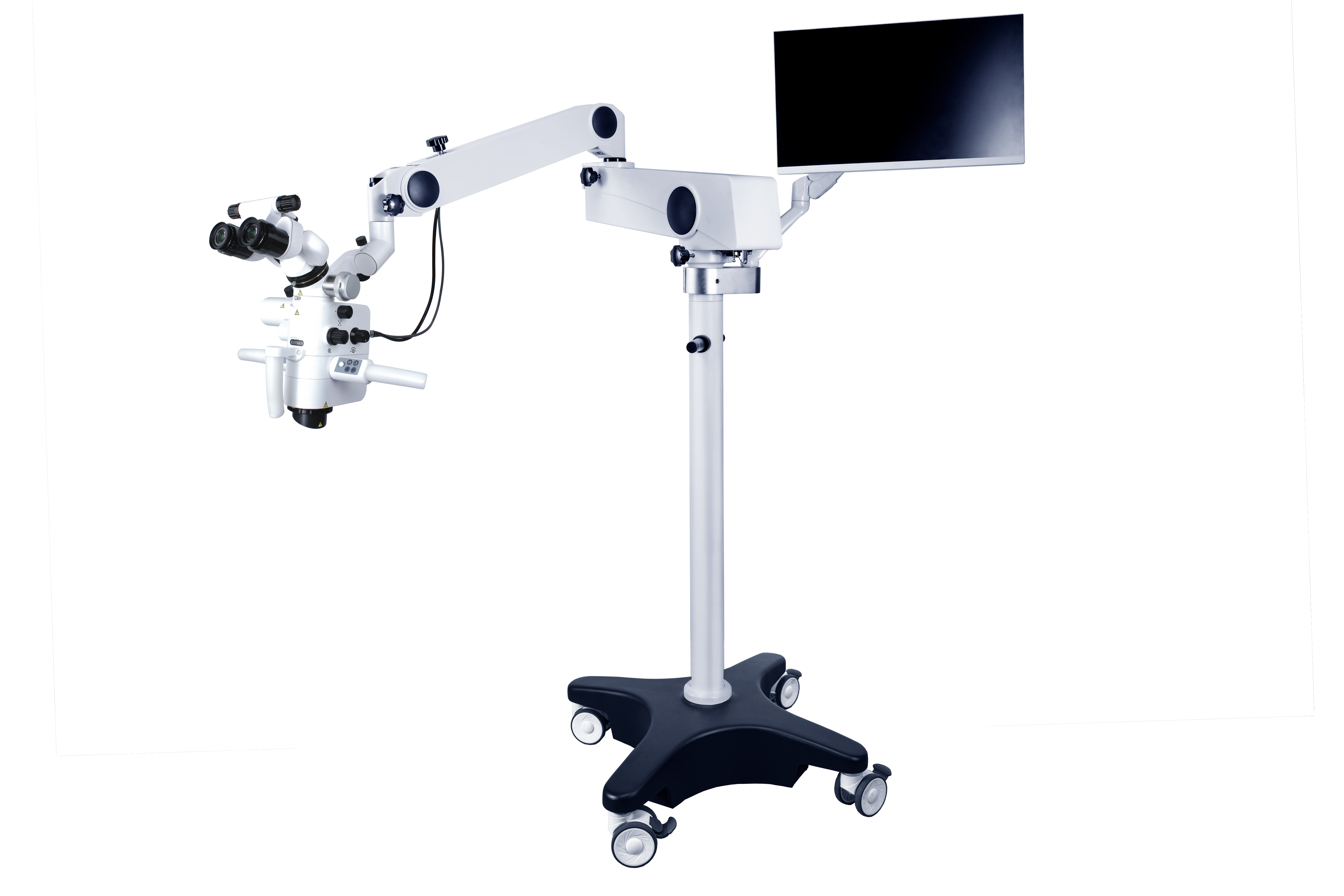
CORDER తీసుకుంటున్నారుASOM-520-D డెంటల్ సర్జికల్ మైక్రోస్కోప్ఉదాహరణకు, ఐపీస్ యొక్క మాగ్నిఫికేషన్ 10 × నుండి 15 × వరకు ఉంటుంది, సాధారణంగా ఉపయోగించే మాగ్నిఫికేషన్ 12.5X, మరియు ఆబ్జెక్టివ్ లెన్స్ యొక్క ఫోకల్ లెంగ్త్ 200~500 మిమీ పరిధిలో ఉంటుంది. మాగ్నిఫికేషన్ ఛేంజర్ రెండు ఆపరేటింగ్ మోడ్లను కలిగి ఉంది: ఎలక్ట్రిక్ స్టెప్లెస్ సర్దుబాటు మరియు మాన్యువల్ నిరంతర మాగ్నిఫికేషన్ సర్దుబాటు.
యొక్క ప్రకాశం వ్యవస్థశస్త్రచికిత్స సూక్ష్మదర్శినిఫైబర్ ఆప్టిక్ లైట్ సోర్స్ ద్వారా అందించబడుతుంది, ఇది వీక్షణ క్షేత్రానికి ప్రకాశవంతమైన సమాంతర ప్రకాశాన్ని అందిస్తుంది మరియు శస్త్రచికిత్స క్షేత్ర ప్రాంతంలో నీడలను ఉత్పత్తి చేయదు. బైనాక్యులర్ లెన్స్లను ఉపయోగించి, రెండు కళ్ళను పరిశీలన కోసం ఉపయోగించవచ్చు, అలసటను తగ్గిస్తుంది; త్రిమితీయ వస్తువు చిత్రాన్ని పొందండి. అసిస్టెంట్ సమస్యను పరిష్కరించడానికి ఒక పద్ధతి ఏమిటంటే అసిస్టెంట్ మిర్రర్ను అమర్చడం, ఇది సర్జన్ మాదిరిగానే స్పష్టమైన వీక్షణను అందించగలదు, కానీ అసిస్టెంట్ మిర్రర్ను అమర్చడానికి అయ్యే ఖర్చు సాపేక్షంగా ఎక్కువగా ఉంటుంది. మరొక పద్ధతి ఏమిటంటే, మైక్రోస్కోప్పై కెమెరా వ్యవస్థను ఇన్స్టాల్ చేయడం, దానిని డిస్ప్లే స్క్రీన్కు కనెక్ట్ చేయడం మరియు సహాయకులు స్క్రీన్పై చూడటానికి అనుమతించడం. బోధన లేదా శాస్త్రీయ పరిశోధన కోసం వైద్య రికార్డులను సేకరించడానికి మొత్తం శస్త్రచికిత్స ప్రక్రియను కూడా ఫోటో తీయవచ్చు లేదా రికార్డ్ చేయవచ్చు.
గుజ్జు మరియు పెరియాపికల్ వ్యాధుల చికిత్స సమయంలో,దంత శస్త్రచికిత్స సూక్ష్మదర్శినిలురూట్ కెనాల్ ఓపెనింగ్లను అన్వేషించడానికి, కాల్సిఫైడ్ రూట్ కెనాల్లను క్లియర్ చేయడానికి, రూట్ కెనాల్ గోడ చిల్లులను సరిచేయడానికి, రూట్ కెనాల్ పదనిర్మాణం మరియు శుభ్రపరిచే ప్రభావాన్ని పరిశీలించడానికి, విరిగిన పరికరాలను మరియు విరిగిన రూట్ కెనాల్ పైల్స్ను తొలగించడానికి మరియుసూక్ష్మ శస్త్రచికిత్సపెరియాపికల్ వ్యాధుల విధానాలు.
సాంప్రదాయ శస్త్రచికిత్సతో పోలిస్తే, మైక్రోసర్జరీ యొక్క ప్రయోజనాలు: రూట్ అపెక్స్ యొక్క ఖచ్చితమైన స్థానం; ఎముక యొక్క సాంప్రదాయ శస్త్రచికిత్స విచ్ఛేదనం పెద్ద పరిధిని కలిగి ఉంటుంది, తరచుగా 10mm కంటే ఎక్కువ లేదా సమానంగా ఉంటుంది, అయితే మైక్రోసర్జికల్ ఎముక విధ్వంసం చిన్న పరిధిని కలిగి ఉంటుంది, 5mm కంటే తక్కువ లేదా సమానంగా ఉంటుంది; సూక్ష్మదర్శినిని ఉపయోగించిన తర్వాత, పంటి మూలం యొక్క ఉపరితల స్వరూపాన్ని సరిగ్గా గమనించవచ్చు మరియు రూట్ కటింగ్ వాలు యొక్క కోణం 10° కంటే తక్కువగా ఉంటుంది, అయితే సాంప్రదాయ రూట్ కటింగ్ వాలు యొక్క కోణం పెద్దది (45°); రూట్ కొన వద్ద రూట్ కెనాల్ల మధ్య ఇస్త్మస్ను గమనించే సామర్థ్యం; రూట్ చిట్కాలను ఖచ్చితంగా సిద్ధం చేసి నింపగలగాలి. అదనంగా, ఇది రూట్ ఫ్రాక్చర్ సైట్ మరియు రూట్ కెనాల్ వ్యవస్థ యొక్క సాధారణ శరీర నిర్మాణ సంబంధమైన మైలురాళ్లను గుర్తించగలదు. క్లినికల్, బోధన లేదా శాస్త్రీయ పరిశోధన ప్రయోజనాల కోసం డేటాను సేకరించడానికి శస్త్రచికిత్స ప్రక్రియను ఫోటోగ్రాఫ్ చేయవచ్చు లేదా రికార్డ్ చేయవచ్చు. దీనిని పరిగణించవచ్చుదంత శస్త్రచికిత్స సూక్ష్మదర్శినిలుదంత గుజ్జు వ్యాధుల నిర్ధారణ, చికిత్స, బోధన మరియు క్లినికల్ పరిశోధనలో మంచి అనువర్తన విలువ మరియు అవకాశాలను కలిగి ఉన్నాయి.
పోస్ట్ సమయం: డిసెంబర్-19-2024


